 WalkitoGet the app
Posterior tibial tendon dysfunction exercises: what helps and when
Posterior tibial tendon dysfunction, often called PTTD, happens when the tendon that supports the arch on the inside of the ankle weakens or becomes inflamed. It is the most common cause of adult-acquired flatfoot. Exercise can help in the early stages, but not every stage responds the same way, and the evidence is still limited. This page covers the stages, the exercises with the best support, and when a boot or surgery enters the picture.
The posterior tibial tendon runs behind the inner ankle bone and attaches under the foot. It holds the arch up during walking. When it degenerates, the arch flattens, the heel tilts outward, and pain settles along the inner ankle or inner midfoot. A 2018 systematic review found only three randomized or controlled trials totalling 93 people, so the exercise evidence here is much thinner than for conditions like plantar fasciitis or Achilles tendonitis.
Walkito turns these exercises into a short daily plan that adjusts to how your feet felt this morning. Get Walkito on the App Store →
What is posterior tibial tendon dysfunction?
The tibialis posterior is a muscle in the deep calf. Its tendon passes behind the inner ankle bone, the medial malleolus, and fans out under the foot to attach to several bones in the midfoot. During walking, it pulls the arch up and controls how much the foot rolls inward. When the tendon degenerates or tears, that support is lost.
PTTD is the most common cause of adult-acquired flatfoot. It is more common in women, in people over 40, and in those with a higher BMI. Risk factors include hypertension, diabetes, and previous steroid injections near the tendon. The pain typically sits behind or below the inner ankle bone and gets worse with activity, especially walking uphill or on uneven ground.
The clinical name you may see is tibialis posterior tendinopathy. Older sources sometimes call it posterior tibial tendonitis when the problem is mainly inflammation, or posterior tibial tendon insufficiency when the tendon has lengthened and the arch has started to drop. All three terms describe the same spectrum.
What are the stages of PTTD?
Clinicians grade PTTD in four stages. The stage determines whether exercise and orthoses are likely to help, or whether a boot, brace, or surgery should be discussed.
Stage I means the tendon is inflamed but still intact. The arch looks normal. There is pain and sometimes mild swelling behind the inner ankle. Strength on a single-leg heel raise is usually reduced compared with the other side, but the shape of the foot has not changed. Exercise and supportive orthoses are the main approach.
Stage II means the tendon has lengthened or partially torn. The arch has dropped and the heel tilts outward, but the deformity is still flexible, meaning a clinician can push the foot back into position. This is the stage most of the exercise research covers. Orthoses, stretching, and progressive strengthening are the standard conservative program.
Stage III means the deformity has become rigid. The foot cannot be pushed back into a corrected position, and there are often arthritic changes in the joints below the ankle. Exercise alone is unlikely to change the shape of the foot at this stage. Surgical assessment is usually discussed.
Stage IV adds ankle joint involvement. The ankle tilts into a valgus position, meaning it leans outward. This is the most advanced stage and typically requires surgical intervention.
Most people who search for PTTD exercises are in stage I or II. If you are not sure which stage applies to you, a clinician can tell you with a physical exam and, if needed, imaging. The single-heel-raise test, where you try to rise onto one foot, is a quick screening tool: difficulty rising, pain during the rise, or the heel not tilting inward at the top all suggest posterior tibial weakness.
What does the exercise evidence say?
A 2018 systematic review identified three randomized controlled trials with 93 participants total. Two tested strengthening on top of orthoses and stretching; the third tested strengthening and balance training against no intervention. The review found moderate effect sizes (SMD 0.6 to 1.2) for eccentric strengthening in reducing pain and disability compared with orthoses and stretching alone.
The largest of the three studies was a 2009 RCT of 36 people with stage I or II PTTD. Participants were assigned to one of three groups for three months: orthoses and stretching only, orthoses plus stretching plus concentric exercise, or orthoses plus stretching plus eccentric exercise. Both exercise groups improved more than the orthoses-only group. The eccentric group achieved loads 3.3 times higher than the concentric group by the end of the program, though both exercise types reduced pain.
A second RCT, published in 2015, randomized 39 people with stage II PTTD to orthoses plus stretching or orthoses plus stretching plus strengthening (including band exercises and heel raises). Both groups improved, but the strengthening group did not separate significantly from the stretching group on the primary outcome. The authors suggested the strengthening dose may not have been high enough.
The review authors concluded that the evidence supports adding progressive resistance exercise to orthoses for early PTTD, but called for larger trials. This is an area where the research is still catching up to clinical practice.
Ross 2018: 3 studies, n = 93. Kulig 2009: n = 36, 3 arms, 3 months, Foot Function Index, pain VAS. Houck 2015: n = 39, 2 arms, 3 months, FAAM.
The exercises
The exercises below target the tibialis posterior muscle and the muscles that support the arch. Band inversion trains the tibialis posterior directly. Heel raises with a focus on arch lift load the tendon during a functional movement. Short foot strengthens the intrinsic muscles that help the arch. Calf stretching addresses the common finding of reduced ankle dorsiflexion in people with PTTD.
The exercise programs in the trials also included calf stretching and orthoses. Orthoses were not optional in any study. If you have PTTD, an arch-supporting orthosis is part of the program, not a substitute for exercise or the other way around.
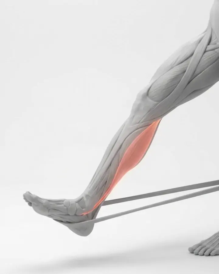
Band inversion (resisted turn-in)
Sit with your legs out in front of you and a resistance band looped around the forefoot. Anchor the other end to a table leg or have someone hold it to the outside. Turn the foot inward against the band, keeping the knee still. The movement comes from the ankle, not the leg.
Evidence: Moderate. Resisted inversion was part of the Kulig 2009 RCT exercise protocol. The tibialis posterior is selectively activated during inversion (Kulig 2004 MRI study).
Sharp pain behind the inner ankle bone
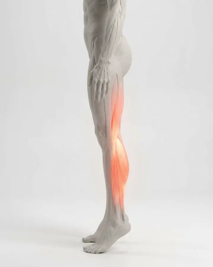
Heel raises (with ball between ankles)
Stand on both feet with a small ball or rolled towel between the inner ankle bones. Rise onto your toes while squeezing the ball inward. The squeeze cues the tibialis posterior to fire. Progress to single-leg raises once the double-leg version is comfortable for two sessions in a row.
Evidence: Moderate. Heel raises with arch engagement were included in the Houck 2015 RCT. MRI shows moderate tibialis posterior activation during heel raises (Kulig 2004).
Pain behind the inner ankle that is above 6/10
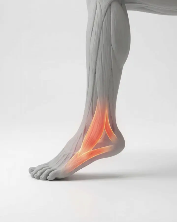
Short foot (seated)
Sit with your foot flat on the floor. Without curling the toes, draw the ball of the foot toward the heel. The arch should rise slightly. This trains the intrinsic foot muscles that work alongside the tibialis posterior to support the arch.
Evidence: Moderate. Short foot exercise was part of the combined program in studies on flat-foot correction. A 2024 review found it improved foot posture in programs longer than six weeks.
Pain in the arch or inner ankle
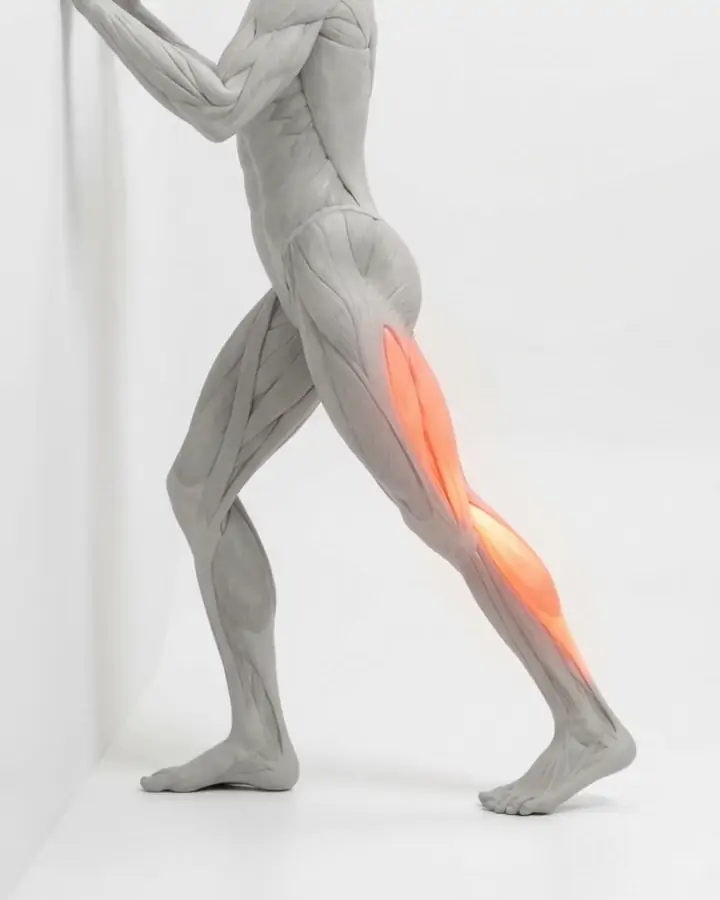
Calf stretch (straight knee)
Hands on a wall. Back leg straight, heel down, hips forward. Hold until you feel the stretch in the upper calf. Reduced ankle dorsiflexion is common in PTTD and can contribute to compensatory pronation.
Evidence: Moderate. Calf stretching was included in all three PTTD exercise trials as a baseline intervention.
Pain in the Achilles tendon
Do orthoses help, and should you wear them during exercise?
Every PTTD exercise trial used orthoses as part of the baseline intervention. Orthoses were not tested against exercise; they were tested with exercise on top. The 2009 RCT found that orthoses and stretching alone improved function, and that adding exercise improved it further.
A rigid or semi-rigid arch-supporting orthosis keeps the arch from collapsing during standing and walking. For the exercises on this page, you can do band inversion and short foot without shoes, but heel raises should be done in the same supportive shoes you walk in, because the arch needs support under load.
Over-the-counter arch supports may be enough for stage I. Custom orthoses are more common for stage II, where the deformity is greater. A clinician or podiatrist can help you decide which is appropriate.
When is a boot or surgery discussed?
A walking boot or ankle brace is sometimes used in stage I or II PTTD when pain is too high for exercise. The boot immobilizes the tendon to let acute inflammation settle. It is a short-term measure, not a long-term program. Once pain drops enough, exercise and orthoses take over.
Surgery is usually discussed when conservative management has not improved symptoms after several months, or when the deformity has progressed to stage III or IV. Surgical options range from tendon repair and transfer in earlier stages to joint fusion in later stages. The decision depends on the stage, the degree of deformity, and the person.
This page does not cover surgical options in detail. If you are in stage III or IV, or if stage II symptoms have not responded to exercise and orthoses over several months, a foot and ankle specialist can discuss next steps.
How does PTTD relate to flat feet?
PTTD is the most common cause of adult-acquired flatfoot, meaning a flat foot that develops in adulthood rather than being present since childhood. If you had normal arches and they have started to drop on one side, with pain behind the inner ankle, PTTD is the most likely explanation.
Congenital flexible flat feet, the kind present since childhood, are a different situation. They may never cause symptoms. The exercises overlap: short foot, band inversion, and heel raises appear in both flat feet exercises and PTTD programs. But the clinical context is different, and PTTD usually needs orthotics and closer monitoring because the tendon is degenerating, not simply lax.
If you are not sure whether your flat foot is long-standing or new, a clinician can compare the two feet, check the tendon, and look at the heel alignment when you stand.
How long does improvement take?
The two RCTs ran for three months. Both showed improvement by the end of the program, but neither tracked participants long-term. A small pilot study of ten patients by the same group also ran for about two and a half months of twice-daily eccentric loading and found symptom improvement along with some tendon changes on ultrasound.
In clinical practice, improvement from stage I or II PTTD with exercise and orthoses is measured in months, not weeks. Tendon degeneration does not reverse quickly. A reasonable expectation is reduced pain and better function over three to six months, with ongoing maintenance exercise after that. If there is no improvement after three months of consistent exercise and orthoses, it is worth going back to a clinician to reassess the stage.
Questions people ask
What exercises help posterior tibial tendon dysfunction?
A 2018 systematic review of three studies with 93 people found that eccentric and concentric strengthening exercises, added to orthoses and stretching, reduced pain and improved function more than orthoses and stretching alone (Ross 2018). The exercises tested include resisted foot inversion with a band, progressive heel raises, and calf stretching.
Can you reverse PTTD with exercise?
In stages I and II, where the tendon is inflamed or lengthened but the foot is still flexible, exercise and orthoses can reduce pain and improve function. No study has shown that exercise reverses tendon degeneration or restores a dropped arch to its original height. In stages III and IV, where the deformity is rigid, exercise alone is unlikely to change the shape of the foot.
What is the difference between PTTD and flat feet?
PTTD is the most common cause of adult-acquired flatfoot, a flat foot that develops in adulthood because the tibialis posterior tendon weakens. Congenital flat feet, present since childhood, are a different situation. They may never cause pain. PTTD usually affects one side, gets worse over time, and comes with pain behind the inner ankle.
What does posterior tibial tendon pain feel like?
The pain usually sits behind or below the inner ankle bone. It can extend along the inner arch. It gets worse with activity, especially walking uphill, on uneven ground, or during single-leg balance. The area may swell. A common early sign is difficulty doing a single-leg heel raise on the affected side.
Do I need orthotics for PTTD?
Every exercise trial for PTTD used orthoses as part of the baseline program. Orthoses support the arch and reduce the load on the tendon during walking. The evidence does not support exercise without orthoses for PTTD. Over-the-counter arch supports may work for mild cases, while custom orthoses are more common for stage II.
Is PTTD the same as inside ankle pain?
PTTD is one of the more common causes of pain on the inner side of the ankle, but it is not the only one. Other possibilities include a deltoid ligament sprain, a navicular stress fracture, or tarsal tunnel syndrome (nerve compression). A clinician can distinguish these with a physical exam. PTTD usually comes with a flattening arch and difficulty with single-leg heel raises.
When does PTTD need surgery?
Surgery is usually discussed when conservative management, meaning exercise, orthoses, and sometimes a walking boot, has not improved symptoms after several months, or when the deformity has progressed to stage III or IV, where the foot is rigidly flat or the ankle joint is affected. Most stage I and II cases respond to conservative care.
See a clinician first if
- the arch on one side has visibly dropped or the heel tilts outward when you stand
- you cannot do a single-leg heel raise on the affected side
- the inner ankle is swollen, red, or warm
- pain came on suddenly after an injury or a twist
- numbness or tingling runs along the inner foot, which can suggest tarsal tunnel syndrome rather than PTTD
- pain does not improve after several weeks of exercise and arch support
- both feet are affected and other joints are swollen or stiff, which can point to inflammatory arthritis
- you have diabetes, reduced feeling in your feet, or poor circulation
Doing it as a plan
Walkito includes band inversion, short foot, and heel raises in its exercise library, and schedules them on strength days as part of an arch and balance program. When you mark the inner ankle or arch on the pain map during a check-in, the session adjusts around that input. The app is built for plantar fasciitis and flat feet. PTTD overlaps with both, but the app does not diagnose PTTD and does not replace orthoses or clinical staging.
You pick 3, 5 or 7 days a week and sessions of 3, 5 or 10 minutes. Every 14 days, a short test checks calf endurance, arch hold, and balance, so you can see what is changing. If you have been diagnosed with PTTD, use the app alongside orthoses and check with your clinician that the stage is appropriate for exercise.
Updated
Walkito is an exercise program. It does not diagnose and does not treat. If pain is sharp, getting worse, or stopping you sleeping, see a clinician.
Start with 3 minutes a day.